High Resolution Microendoscopy for the Management of Esophageal Neoplasia & Augmented Reality
Funding Agency:
National Cancer Institute, National Institutes of Health
Collaborators:
Rebecca Richards-Kortum (Rice University), Sharmila Anandasabapathy (Baylor College of Medicine), Chin Hur (Massachusetts General Hospital), Kalpesh Patel (Baylor College of Medicine), Sanford Dawsey (National Cancer Institute), Daniel Rosen (Baylor College of Medicine), Hong Xu (First Hospital of Jilin University, Changchun, China), Fan Zhang (First Hospital of Jilin University, Changchun, China), Guiqi Wang (CICAMS, Beijing, China), Xinying Yu (CICAMS, Beijing, China), William Buras (Tietronix Software Inc)
Overview:
Esophageal cancer is the 6th most common cause of cancer-related mortality worldwide. While the disease carries a significant burden in the United States, certain geographic regions (northern China, east Africa, Iran) have incidence rates >1/1000 and screening protocols have been implemented with limited success. The most widely used method of endoscopic evaluation uses Lugol’s iodine mucosal staining with targeted biopsy of abnormal (unstained) areas. While Lugol’s increases the sensitivity of standard white-light endoscopy, specificity remains poor as inflammation and other benign mucosal change can mimic neoplasia. As a result, many unnecessary biopsies are obtained, increasing time, risk and cost. High-resolution imaging with confocal endomicroscopy dramatically enhances the diagnostic accuracy and yield of Lugol’s and other widefield techniques. However, existing platforms are costly (>$150,000) and only available in a handful of tertiary centers worldwide.
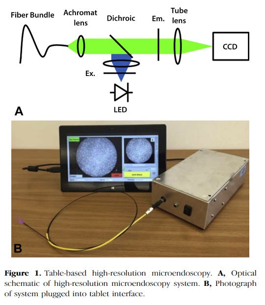
In this study, we are optimizing the High Resolution Microendoscope (HRME) so that it can be used by less experienced users in low-resource areas. We hypothesize that this novel, low cost approach can improve the efficiency, clinical impact and cost-effectiveness of the current standard of esophageal squamous cell neoplasia (ESCN) screening by offering real-time, in vivo diagnosis that reduces biopsy number and repeat procedures while preserving sensitivity. Aims for this study are: (1) develop and optimize a low-cost, mobile HRME platform that be used in low-resource settings by less experienced clinicians, (2) perform a prospective, randomized clinical trial comparing the current standard of Lugol’s chromoendoscopy to our proposed ‘optical biopsy’ approach in 1300 subjects in China and the United States, and (3) construct, refine, and analyze a disease model of ESCN.
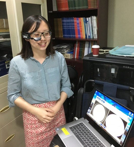
In addition, we were funded for a supplement in 2016 to refine and validate a more mobile, low-cost Augmented Reality (AR) platform to facilitate real-time endoscopic screening by non-expert clinicians worldwide (and proposed pilot sites for evaluation in Year 5, e.g., South Africa). Using AR glasses and a small tablet computer, microendoscopic images with a quantitative software interpretation (neoplasia vs. no neoplasia) will be displayed in real-time colocalized with white light endoscopic images, allowing easier and more accurate biopsy and treatment. Revised aims are: (1) Adapt our mobile HRME platform to provide an automated diagnosis in real-time (marHRME), with colocalization of both the microendoscopic and endoscopic images for more accurate biopsy and treatment, (2) evaluate whether the marHRME alters the endoscopist’s approach (biopsy, not biopsy, treat at the point of care) and leads to a reduction in biopsy number and repeat procedures, (3) incorporate the HRME and marHRME into endoscopic screening/surveillance programs in different global settings.
Publications under current grant funding:
Quang T, Schwarz RA, Dawsey SM, et al. A tablet-interfaced high-resolution microendoscope with automated image interpretation for real-time evaluation of esophageal squamous cell neoplasia. Gastrointestinal endoscopy. 2016;84(5):834-841. doi:10.1016/j.gie.2016.03.1472.
Related recent publications under previous grant funding:
Shin D, Lee MH, Polydorides AD, Pierce MC, Vila PM, Parikh ND, Rosen DG, Anandasabapathy S and Richards-Kortum R. Quantitative analysis of high-resolution microendoscopic images for diagnosis of neoplasia in patiens with Barret’s esophagus. Gastrointest Endosc (2016);83(1):107-114.
Protano MA, Xu H, Wang G, Polydorides AD, Dawsey SM, Cui J, Xue L, Zhang F, Quang T, Pierce MC, Shin D, Schwarz RA, Bhutani MS, Lee M, Parikh N, Hur C, Xu W, Moshier E, Godbold J, Mitcham J, Hudson C, Richards-Kortum RR and Anandasabapathy S. Low-cost high-resolution microendoscopy for the detection of esophageal squamous cell neoplasia: an international trial. Gastroenterol 2015;149(2): 321-329.
Shin D, Protano MA, Polydorides AD, Dawsey SM, Pierce MC, Kim MK, Schwarz RA, Quang T, Parikh N, Bhutani MS, Zhang F, Wang G, Xue L, Wang X, Xu H, Anandasabapathy S and Richards-Kortum R. Quantitative analysis of high resolution microendoscopic images for diagnosis of esophageal squamous cell carcinoma. Clin Gastroenterol Hepatol (2015);13:272-279.
Ishijima A, Schwarz RA, Shin D, Mondrik S, Vigneswaran N, Gillenwater AM, Anandasabapathy S and Richards-Kortum R. Automated frame selection process for high-resolution microendoscopy. J Biomed Opt (2015);20(4):046014.
Pictures:
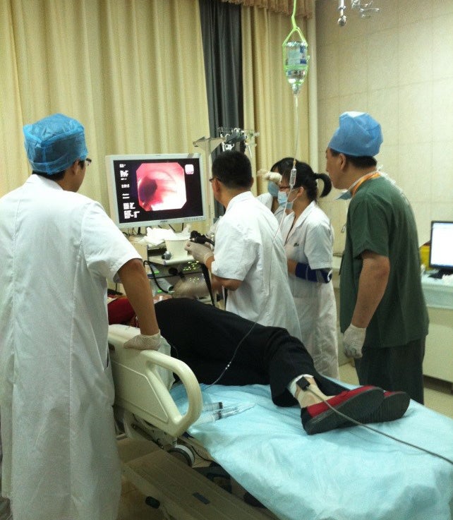
Clinical study of endoscopy and HRME imaging in collaboration with the Cancer Institute at the Chinese Academy of Medical Sciences (CICAMS), Beijing, China.