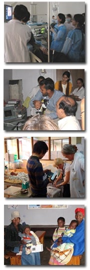
Rebecca Richards-Kortum is the Malcolm Gillis University Professor and a member of the Department of Bioengineering and the Department of Electrical & Computer Engineering at Rice University in Houston, Texas. Dr. Richards-Kortum’s research has been instrumental in improving early detection of cancer and other diseases, especially in low-resource settings. Her Optical Spectroscopy and Imaging Laboratory integrates advances in nanotechnology and molecular imaging with microfabrication technologies to develop optical imaging systems that are inexpensive, portable, and provide point-of-care diagnosis.
The Kortum Lab works closely with clinical collaborators to develop multi-scale, multi-modal optical technologies for cancer screening and diagnosis in organ sites including the oral cavity, esophagus, stomach, cervix, and bladder. One of the lab’s most versatile technologies is the high-resolution microendoscope (HRME), a low-cost fiber-optic fluorescence microscope that can image the size, shape, and distribution of epithelial cell nuclei in real time in vivo, delineating morphologic changes associated with dysplasia and cancer. The HRME is often used in combination with the lab’s other imaging technologies that identify high-risk areas, such as autofluorescence imaging or vital-dye fluorescence imaging. Other technologies with high spatial or spectral resolution include high-resolution microvasculature imaging, structured illumination microscopy, and fluorescence & reflectance spectroscopy. For esophageal cancer screening, we are developing a swallowable tethered capsule with peristalsis-driven rotation capable of imaging the entire esophagus. For cervical cancer screening, we have developed a paper-based human papillomavirus (HPV) DNA test that is easy to use, runs within an hour, has equivalent sensitivity to gold standard assays, and costs $2-3 per test.
We evaluate our cancer screening technologies through translational clinical studies in a variety of high-resource and low-resource health care settings. We have ongoing cervical cancer imaging studies in collaboration with M.D. Anderson Cancer Center in Houston, the Instituto del Cáncer de El Salvador, and Barretos Cancer Hospital in Barretos, Brazil. We carry out esophageal imaging studies in collaboration with Baylor College of Medicine in Houston, the Cancer Institute at the Chinese Academy of Medical Sciences, and Jilin University in China. We have done gastric imaging studies in Honduras, and we have ongoing oral and bladder imaging studies at the Texas Medical Center in Houston. We are evaluating our paper-based HPV DNA test in El Salvador. We also collaborate with M.D. Anderson Cancer Center to develop models that can be used to train health care providers in low-resource settings to perform cervical cancer screening and treatment.
Funded Projects
- Low Cost Tethered Capsule Endoscope with High-Resolution Digital Chromoscopy for Barrett’s Screening
- Precision Optical Guidance for Oral Biopsy Based on Next-Generation Hallmarks of Cancer
- Low-Cost Mobile colposcopy and confocal imaging for global prevention of cervical cancer
- The Effectiveness of High Resolution Microendoscopy (HRME) in High Grade Intraepithelial Lesions (HSIL) Diagnosis for People Living with HIV
- Deep Learning Microscope For Slide-Free and Digital Histology
- A Sample-to-Answer Point-of-Care Diagnostic for Recently Transfused Sickle Cell Anemia Patients in Low Resource Settings
- The AVANCO Research Consortium: A Mozambique/Brazil/Texas Alliance to advance novel and affordable technologies for the prevention and diagnosis of cervical cancer of women living with HIV
- The Center for Innovation and Translation of Point of Care Technologies for Expanded Cancer Care Access (CITEC)
- AI-Assisted Microendoscopy for the Early Detection of Esophageal Cancer
- AccessPath: A Low Cost Rugged End to End System for Slide Free Pathology Based on Computational Microscopy and Pan Cancer Targeted Vital Dyes
- Sample-to-answer HPV DNA nucleic acid test for cervical cancer screening in Mozambique
